Harnessing immune complexity.
The extensive immunology and inflammation experience at The University of Manchester has been drawn together into a multidisciplinary research institute – The Lydia Becker Institute of Immunology and Inflammation.
We unite basic, translational, and clinical research to address the complex and ever-increasing role immunology plays in modern medicine.
Research
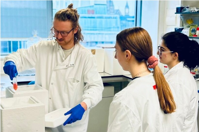
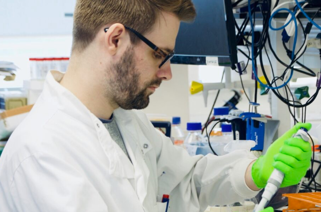
Our research
We remove traditional boundaries to find solutions for today’s global disease challenges.
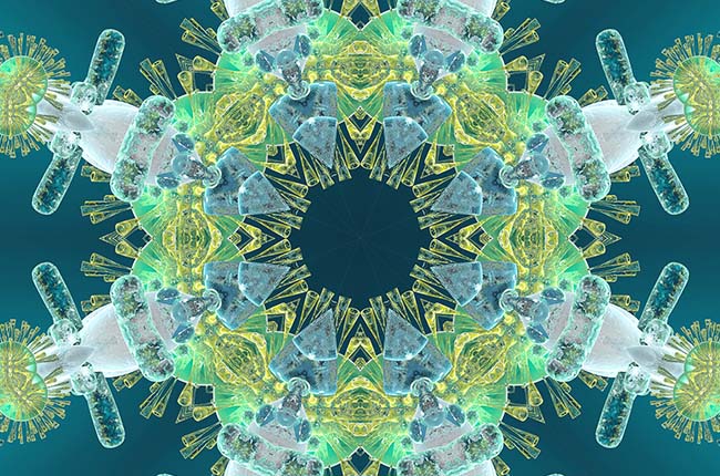
COVID-19 activity
We’re uniquely placed to understand how COVID-19 behaves and affects individuals.
More about our Institute


Contact us
Please get in touch if you’d like to know more about what we do.
Institute Director
Professor Tracy Hussell
Email: tracy.hussell@manchester.ac.uk
Institute Manager
Mr Ameur Bayar
Email: ameur.bayar@manchester.ac.uk
Tel: +44 (0)161 306 3771
Follow us
Twitter: @LydiaBeckerIII
YouTube: Lydia Becker Institute